ScanNav Anatomy PNB will help tip the balance of safety and confidence in favour of performing regional anaesthesia. Our aim is to make a real clinical difference to patients by increasing the availability of regional anaesthesia through cutting edge technology
Dr David Burckett-St.Laurent, Consultant Anaesthetist, Aneurin Bevan University Health Board
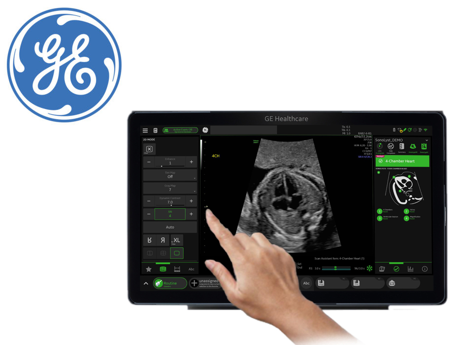
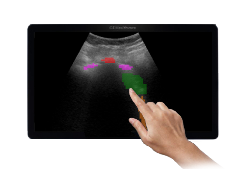
ScanNav Anatomy uses the latest AI technology to automatically highlight the live ultrasound image and make it easier to identify key anatomical structures. It supports the performance of healthcare professionals who are suitably qualified, but who perform ultrasound-guided procedures on a less frequent basis. It also enhances the accuracy and standardisation of ultrasound image interpretation by making it easier to identify key anatomical structures.
ScanNav Anatomy Peripheral Nerve Block
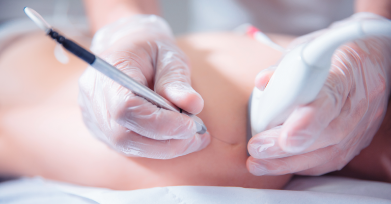
Ultrasound-guided peripheral nerve blocks are increasingly being used as a prudent alternative to general anesthesia, but not all anesthetists have the specialist knowledge of ultrasound anatomy to perform them.
Available in the UK as a stand-alone device, mounted on a portable cart that can be plugged into existing anesthesiology ultrasound machines, ScanNav Anatomy Peripheral Nerve Block supports nine common peripheral nerve blocks. Clinicians get continuous feedback from real-time highlighting of key anatomical structures relevant to the block on their live ultrasound. The system’s integrated reference material, including 3D animations relating the ultrasound view to the underlying anatomy, allows users to re-familiarize themselves with blocks that they carry out less frequently.
Through the adoption of ScanNav Anatomy PNB, it is hoped that hospitals will be able to increase the number of ultrasound-guided nerve blocks that they can perform.
ScanNav Anatomy has also been submitted for FDA regulatory approval and as such is not available for sale in the US or any other territory requiring government approval for this type of product. Outside the UK and Europe, this material should be considered informational only and does not constitute an offer to sell or infer claims or benefits.
9 Supported Procedures
ScanNav Anatomy PNB supports the teaching of the following nine popular ultrasound guided regional anesthesia procedures:
- Interscalene
- Superior Trunk
- Supraclavicular
- Axillary
- Erector Spinae Plane
- Rectus Sheath
- Suprainguinal Fascia Iliaca
- Adductor Canal / Sub-sartorial femoral triangle
- Popliteal
Key Features
- Compatible with general purpose ultrasound machines with HDMI or DVI second monitor port
- Highlights the key structures relevant to each procedure in real-time
- Complete with block-specific 3D-animated training material
- Can be cleaned with medical grade disinfectants and touchscreen can be operated while wearing gloves
Clinical validation
In a validation study regional anesthesia experts judged the AI-driven colour highlighting to be helpful:
- for identifying anatomical structures in 1,330/1,334 cases (99.7%)
- for confirming the correct ultrasound view in 273/275 ultrasound scans (99.3%)
ScanNav Anatomy PNB Trainer
ScanNav Anatomy PNB is also available as a training simulator for medical learning on volunteers, prior to patient contact.
Dr. David Burckett-St.Laurent
Consultant Anaesthetist, Aneurin Bevan Health Board
Clinical papers for reference
Bowness J, El-Boghdadly K, Burckett-St Laurent D. Artificial intelligence for image interpretation in ultrasound-guided regional anaesthesia. Anaesthesia. 2020 Jul 29. doi: 10.1111/anae.15212. Epub ahead of print. PMID: 32726498.
Turbitt LR, Mariano ER, El-Boghdadly K. Future directions in regional anaesthesia: not just for the cognoscenti. Anaesthesia. 2020 Mar;75(3):293-297. doi: 10.1111/anae.14768. Epub 2019 Jul 3. PMID: 31268173.
Automated image analysis
for protocol-based scanning.
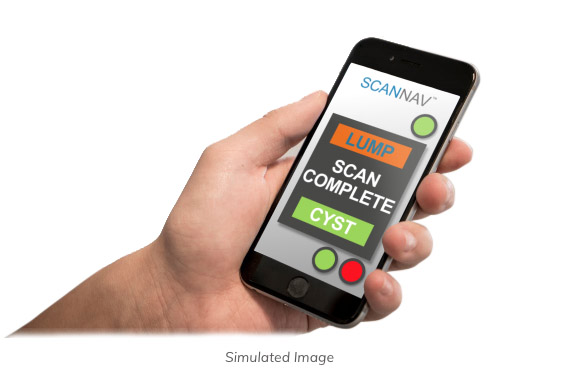
Automated pathology highlighting
for triaging patients.
Automated health check assessments
for the worried well at home.